
Retinal imaging
Fundus photography
A clearer record of the back of your eye.
Fundus photography captures images of the retina and its blood vessels. These pictures help document findings and support conversations about your eye health.
Your branch, schedule, and preparation will be confirmed by our team.
CAM SUR EYE CENTER
Care, explained one step at a time.
Retinal photographs
A visual record for follow-up
Before your visit
Ask about preparation and total visit time.
Practical details
Confirm branch, price, and coverage.
Is this for you?
When might your doctor recommend it?
- Your doctor wants to document the appearance of your retina.
- You need monitoring of an existing retinal condition.
- Your care team wants images to compare with future examinations.
Not sure which test you need? Start with an eye consultation. A test is selected to answer a clinical question; it is not automatically needed for every patient.
What to expect
Three steps, with guidance along the way.
01
Get ready
Ask whether your pupils will need dilation. If so, plan for temporary blur and light sensitivity, and arrange transport as advised.
02
Have the test
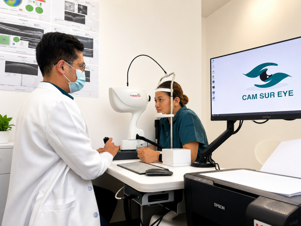
Position your head at the camera and look where the operator asks while photographs are taken.
03
Understand the next step
Ask when your results will be reviewed, who will explain them, and whether another appointment is needed. Bring your questions to that discussion.
The test, explained
About your test.
What it is. Fundus photography produces images of the back of your eye, including the retina, its blood vessels, and the optic nerve head. These images provide a record that your doctor can examine and compare with later photographs. They support your assessment alongside an eye examination and any other recommended tests.
How it works. A specialised retinal camera uses light and optical lenses to photograph the back of the eye through the pupil. You rest your head on the supports and look in the directions the operator asks. Dilating drops may be used to improve the view, and the camera’s light can briefly feel dazzling.
How to prepare. Bring your test request and previous eye records if available, and ask whether dilation is planned. If drops will be used, bring sunglasses and arrange transport home because your vision may be temporarily blurred and bright light may feel uncomfortable. Tell the team about any previous reaction to eye drops.
People behind your care
Support from an eye care team.

YOUR CLINICAL TEAM
Dr. Mark Linus P. Meneses
Medical Director · Ophthalmologist
Learn about the clinic’s medical team, and ask who will perform your test and review your results.
FROM OUR PATIENTS
Get a feel for the clinic.
Hear about the clinic experience directly from patients.
Your questions, answered
A little clarity before you come in.
Does this test hurt?
The camera uses bright light, which can feel dazzling. Tell the team if you are uncomfortable.
How long should I allow for my appointment?
The full visit may include registration, preparation, the test, and review. Ask your branch for the expected total time and whether results will be discussed that day.
Do I need a referral?
Tell our team whether another doctor requested the test, and bring the request and previous results. If you have not been assessed, ask whether a consultation is needed before booking.
How much does it cost, and can I use insurance?
Ask for the current test price and whether consultation or other fees are separate. Give the clinic your provider and plan details so participation, authorization, and any patient share can be checked.
Does this replace a full eye examination?
No. Photographs are one part of an assessment. Your doctor may also recommend an examination or other tests.
Take the next step
Let’s plan your visit.
Send a request for your Fundus photography. Our team will help confirm your branch, the available schedule, and any preparation needed.
Prefer to speak with us? Call your branch.
Nabua
Naga City